Contrast Sensitivity and the Contrast Sensitivity Function
Another important dimension in the measurement of visual function is contrast sensitivity—the sensitivity of an observer to differences in luminance between an object and the background. In general, the higher the contrast, the easier an optotype is to decipher. Over a broad range, the visual system is relatively insensitive to the absolute brightness of a visual stimulus, but is much more attuned to the contrast between adjacent surfaces. For example, the dark ink on a printed page reflects about 10% of the incident light. In comparison, the white paper background has a reflectance of perhaps 90%, regardless of the level of absolute illumination. Thus, when reading under bright sunlight, we still appreciate the printed text as black, even though the absolute brightness of the reflected light is greater than that reflected from white paper in dim illumination, as in twilight. If the brightness of an object (Imin) and the brightness of its background (Imax) are known, the following formula can be used to measure the degree of contrast between the object and its background:
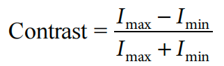
Thus, for typical printed matter, the contrast is about 80% (90% − 10%)/(90% + 10%). Snellen visual acuity is commonly tested with illuminated or projected charts that approximate 100% contrast. Therefore, when we measure Snellen visual acuity, we are measuring, at approximately 100% contrast, the smallest optotype that the visual system can recognize. In everyday life, however, 100% contrast is rarely encountered, and most visual tasks must be performed in lower-contrast conditions.
To take contrast sensitivity into account when measuring visual function, we can use the modulation transfer function (MTF). Consider a target in which the light intensity varies from some peak value to zero in a sinusoidal fashion. The contrast is 100%, but instead of looking like a bar graph, it looks like a bar graph with softened edges. The number of light bands per unit length or per unit angle is called the spatial frequency and is closely related to Snellen acuity. For example, the 20/20 E optotype is composed of bands of light and dark, in which each band is 1 arcmin. Thus, for a target at 100% contrast, 20/20 Snellen acuity corresponds roughly to 30 cycles per degree of resolution when expressed in spatial frequency notation. The relationship between spatial frequency and the contrast sensitivity at each spatial frequency constitutes the MTF.
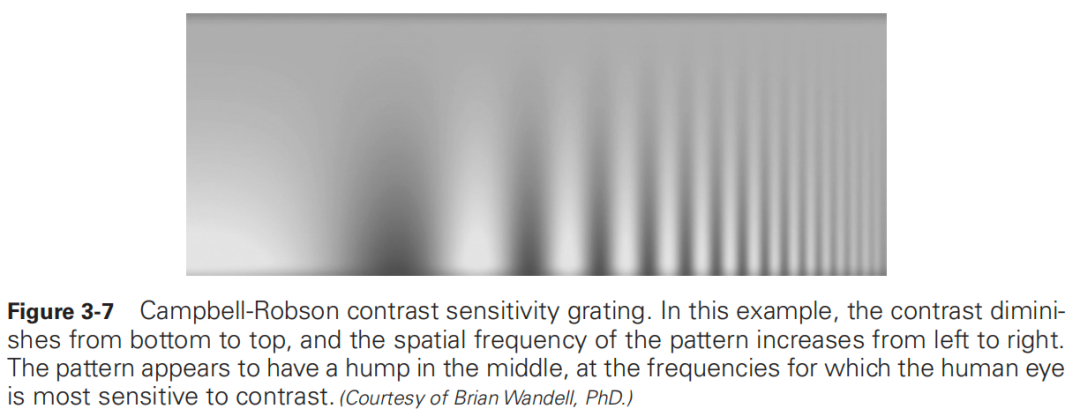
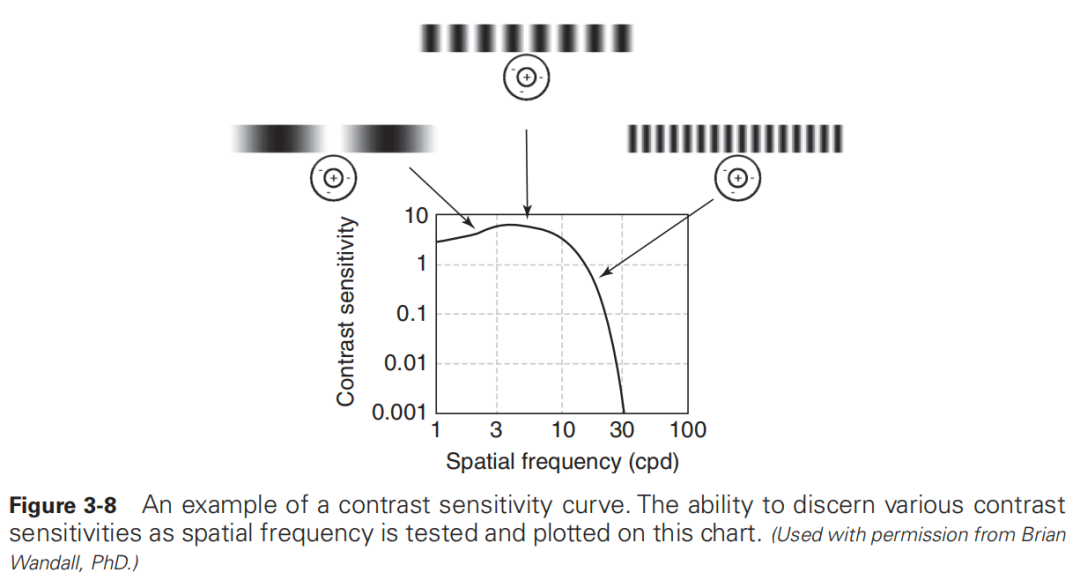
In clinical practice, the ophthalmologist presents a patient with targets of various spatial frequencies and peak contrasts. A plot is then made of the minimum resolvable contrast target that can be seen for each spatial frequency. The minimum resolvable contrast is the contrast threshold. The reciprocal of the contrast threshold is defined as the contrast sensitivity, and the manner in which contrast sensitivity changes as a function of the spatial frequency of the targets is called the contrast sensitivity function (CSF) (Fig 3-7). Figure 3-8 shows a typical contrast sensitivity curve obtained with sinusoidal gratings. Contrast sensitivity can also be tested with optotypes of variable contrast (eg, the Pelli-Robson or Regan charts), which may be easier for patients to use. It is important to perform contrast sensitivity testing with the best possible optical correction in place. In addition, luminance must be kept constant when CSF is tested, because mean luminance affects the shape of the normal CSF curve. In low luminance, the low spatial frequency fall-off disappears and the peak shifts toward the lower frequencies. In brighter light, there is little change in the shape of the normal CSF curve through a range of luminance for the higher spatial frequencies. Generally, contrast sensitivity is measured at normal room illumination, which is approximately 30–70 lux.
Various physiologic and pathologic conditions of the eye affect contrast sensitivity. Any corneal pathology that causes distortion or edema can affect contrast sensitivity. Lens changes, particularly incipient cataracts, may significantly decrease CSF, even with normal Snellen visual acuity. Retinal pathology may affect contrast sensitivity more (as with retinitis pigmentosa or central serous retinopathy) or less (certain macular degenerations) than it does Snellen visual acuity. Glaucoma may produce a significant loss in the midrange of spatial frequencies. Optic neuritis may also be associated with a notch-type pattern of sensitivity loss. Amblyopia is associated with a generalized attenuation of the curve. Pupil size also has an effect on contrast sensitivity. With miotic pupils, diffraction reduces contrast sensitivity; with large pupils, optical aberrations may interfere with performance.
Impairments in perception of contrast may be disqualifying in certain occupational situations, such as driving heavy vehicles. Recognition of these difficulties is often valuable in understanding the concerns of patients, who may have difficulty with certain visual tasks notwithstanding good Snellen acuity as measured with the standard high-contrast eye charts.
In considering the refractive state of the eye, we can use either of the following approaches:
1. The focal point concept: The location of the image formed by an object at optical infinity through a nonaccomodating eye determines the eye’s refractive state. Objects that focus at points anterior or posterior to the retina form blurred images on the retina, whereas objects that focus on the retina form sharp images.
2. The far point concept: The far point is the point in space that is conjugate to the fovea of the nonaccommodating eye; that is, the far point is where the fovea would be imaged if the light rays were reversed and the fovea became the object.
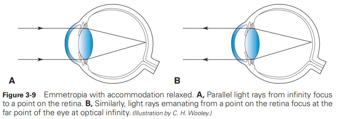
Emmetropia is the refractive state in which parallel rays of light from a distant object are brought to focus on the retina in the nonaccommodating eye (Fig 3-9A). The far point of the emmetropic eye is at infinity, and infinity is conjugate with the retina (Fig 3-9B). Ametropia refers to the absence of emmetropia and can be classified by presumptive etiology as axial or refractive. In axial ametropia, the eyeball is either unusually long (myopia) or short (hyperopia). In refractive ametropia, the length of the eye is statistically normal, but the refractive power of the eye (cornea and/or lens) is abnormal, being either excessive (myopia) or deficient (hyperopia). Aphakia is an example of extreme refractive hyperopia unless the eye was highly myopic (>20.00 D) before lens removal. An ametropic eye requires either a diverging or a converging lens to image a distant object on the retina.
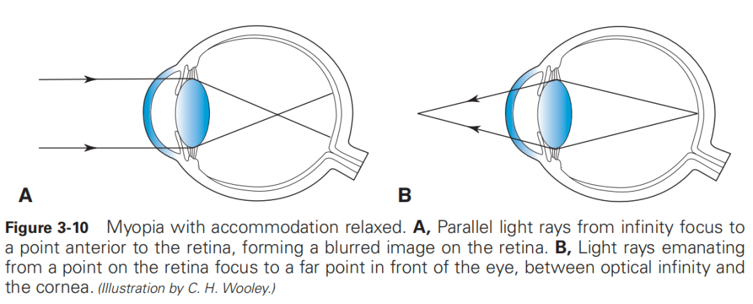
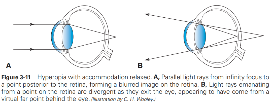
Ametropias may also be classified by the nature of the mismatch between the optical power and length of the eye. In myopia, the eye possesses too much optical power for its axial length, and (with accommodation relaxed) light rays from an object at infinity converge too soon and thus focus in front of the retina (Fig 3-10A). This results in a defocused image on the retina; the far point of the eye is located in front of the eye, between the cornea and optical infinity (Fig 3-10B). In hyperopia, the eye does not possess enough optical power for its axial length, and (with accommodation relaxed) an object at infinity comes to a focus behind the retina, again producing a defocused image on the retina (Fig 3-11A); the far point of the eye (actually a virtual point rather than a real point in space) is located behind the retina (Fig 3-11B).
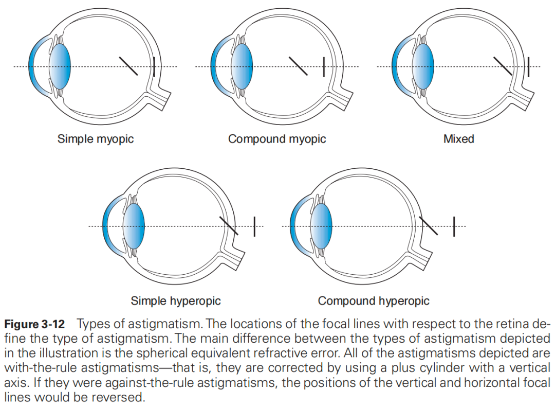
Astigmatism (a =without, stigmos = point) is an optical condition of the eye in which light rays from a point source on the eye’s visual axis do not focus to a single point. Typically, light rays from a single object point are refracted to form 2 focal lines, perpendicular to each other. Each astigmatic eye can be classified by the orientations and relative positions of these focal lines (Fig 3-12). If 1 focal line lies in front of the retina and the other is on the retina, the condition is classified as simple myopic astigmatism. If both focal lines lie in front of the retina, the condition is classified as compound myopic astigmatism. If, in a nonaccommodating eye, 1 focal line lies behind the retina and the other is on the retina, the astigmatism is classified as simple hyperopic astigmatism.If both focal lines lie behind the retina, the astigmatism is classified as compound hyperopic astigmatism. If, in a nonaccommodating eye, one focal line lies in front of the retina and the other behind it, the condition is classified as mixed astigmatism. The orientations of the focal lines reflect, in turn, the strongest and weakest meridians of the net refracting power of the anterior segment refracting surfaces (the cornea and lens). These are referred to as the principal axes.
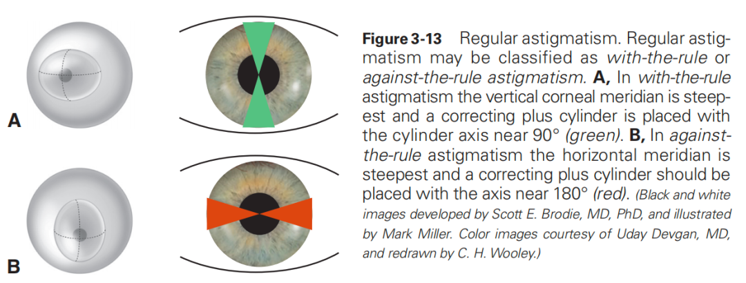
If the principal axes of astigmatism have constant orientation at every point across the pupil, and if the amount of astigmatism is the same at every point, the refractive condition is known as regular astigmatism. Many cases of regular astigmatism may be classified as with-the-rule or against-the-rule astigmatism. In with-the-rule astigmatism (the more common type in children), the vertical corneal meridian is steepest (resembling an American football or a rugby ball lying on its side), and a correcting plus cylinder is placed with the cylinder axis near 90°. In against-the-rule astigmatism (the more common type in older adults), the horizontal meridian is steepest (resembling a football standing on its end), and a correcting plus cylinder should be placed with the axis near 180°. Alternatively, minus cylinders may be placed in the orthogonal directions. The term oblique astigmatism is used to describe regular astigmatism in which the principal meridians do not lie at, or close to, 90° or 180°, but instead lie nearer 45° or 135° (Fig 3-13).
In irregular astigmatism, the orientation of the principal meridians or the amount of astigmatism changes from point to point across the pupil. Although the principal meridians are 90° apart at every point, it may sometimes appear with retinoscopy or keratometry that the principal meridians of the cornea, as a whole, are not perpendicular to one another. Most eyes have at least a small amount of irregular astigmatism, and instruments such as corneal topographers and wavefront aberrometers can be used to detect this condition clinically. These higher-order aberrations in the refractive properties of the cornea and lens may be characterized by Zernike polynomials, which are mathematical shapes that approximate various types of irregular astigmatism more closely than the simple “football” model. These aberrations include such shapes as spherical aberration, coma, and trefoil. See Chapters 1 and 7 of this book and BCSC Section 13, Refractive Surgery, for further discussion.
来源:美国眼科学会(American Academy of Ophthalmology,AAO)2022-2023 Basic and Clinical Science Course ——Clinical Optics and Vision Rehabilitation
本栏目旨在为大家呈现经典眼科教科书,全年365天不停更,非常适合眼科医生和硕士生、博士生自学之用。
当前书籍为《2022-2023基础和临床科学课程》(2022-2023 Basic and Clinical Science Course ,BCSC),是由世界上最大的眼科医师和外科医生协会美国眼科学会(American Academy of Ophthalmology,AAO)组织编写的系列权威课程。
第1节:普通医学更新;
第2节:眼科基础和原理;
第3节:临床光学;
第4节:眼科病理学和眼内肿瘤;
第5节:神经眼科;
第6节:儿科眼科和斜视;
第7节:眼面部整形和眼眶手术;
第8节:外部疾病和角膜;
第9节:葡萄膜炎和眼部炎症;
第10节:青光眼;
第11节:晶状体和白内障;
第12节:视网膜和玻璃体;
第13节:屈光手术
【AI教科书】栏目推出以来,受到了很多同学的喜欢和关注。有不少同学提出,希望可以对每日学习的文稿进行翻译。本着专业负责的态度,我们暂时不会提供AI翻译文本。为大家找到了一些比较靠谱的免费翻译网站,希望可以有所帮助:
知网翻译助手:https://dict.cnki.net/
谷歌翻译:https://translate.google.cn/
有道翻译:https://fanyi.youdao.com/
翻译官:https://fanyi.qq.com/
126期:人眼光学——调节和老视、眼球发育及屈光不正、发育性近视、近视防控、发育性远视
想要获取英文原文PDF、了解更多学习内容,欢迎入群~👇